


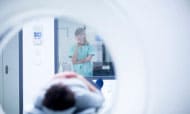
Radiology
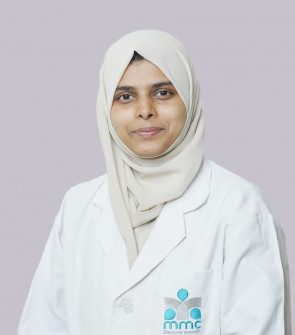
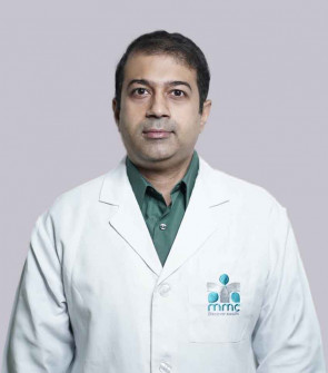
Doctors

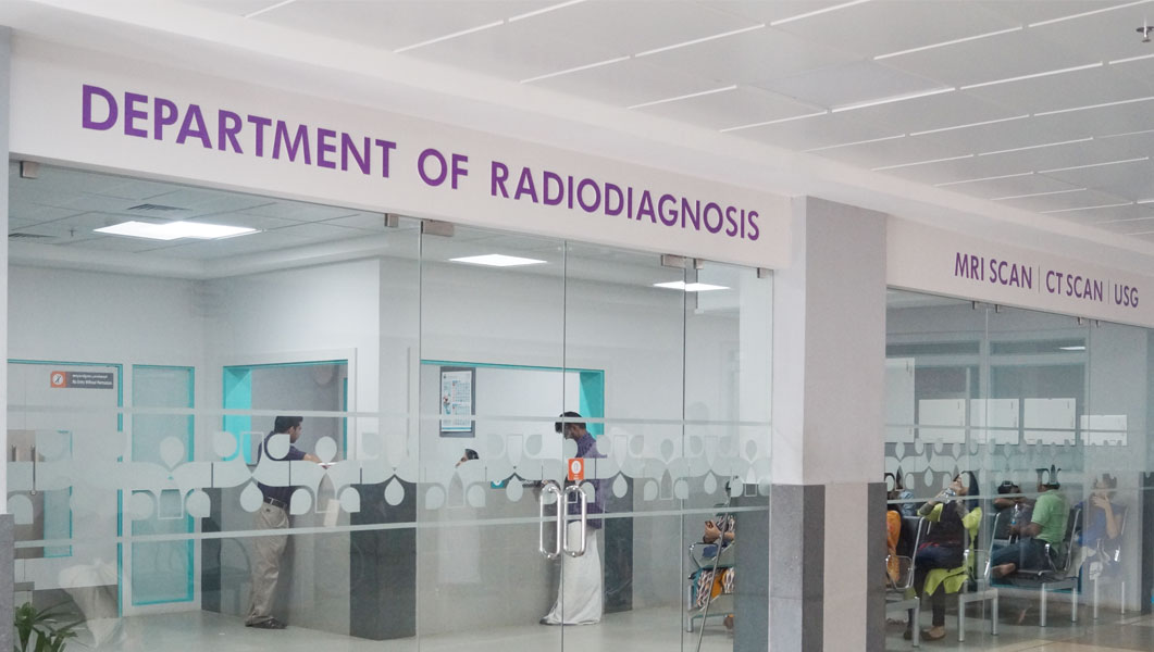
About the Department
Our Radiology Department has a set of expert and experienced Radiologists of Kerala. The department findings helps the doctors in finding out the anomalies in human body with improved perfection.
Our Radio Diagnosis equipments CT Scan and MRI Scan Machines are of the most advanced technology and first of their kind in Kozhikode and in Kerala.
Clinical Focus
Radiology Department of Malabar Medical College focuses on improved diagnosis of the diseases with much more perfection than it was earlier.
- Eliminate the need for exploratory surgery.
- Used to determine when a patient needs surgery.
- Assists in making a diagnosis and further management of most body conditions.
- Interventional radiology, which involves treatment as well as diagnosis, involves less risk, a shorter recovery time and less time in hospital than open surgery or key-hole (laparoscopic) surgery.
- Used to visually guide the treatment of conditions such as heart disease and stroke.
- Used in screening for diseases such as breast cancer (mammography), with early detection reducing the mortality rate.
- Improves cancer diagnosis and is also an effective treatment for cancer and other diseases (known as radiation oncology or radiation therapy).
Infrastructure
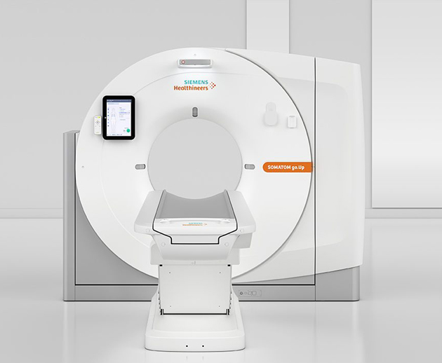
1. CT Scan – 64 Slice
We posses one of the most modern CT Scan equipment with all the facilities to provide maximum comfort to the patient during the imaging processes. Built around a new mobile workflow, SOMATOM go.Up® features a line-up of innovative solutions that bring an unparalleled level of flexibility and mobility to daily CT routines. The solutions also help to enhance patient comfort for potentially higher levels of patient satisfaction.
Features
- First of its kind in Kozhikode and in Kerala.
- Hybrid Technology.
- Built on a unique concept of mobile operationand workflow automation.
- Twin Filter Technology.
- Stellar Detctor Technology.
- CT with Calcium Scoring.
- CT Angio.
- CT Urogram, Virtual colonoscopy, Whole Body CT Scan.
- Brain, Orbit, PNS, Chest, Abdomen & pelvis.
- Spine and Joints with 3D CT.
- Dental CT.
- Temporal Bone CT.
Tablet
- Lightweight, high-resolution tablet gives you total freedom over how you work.
- With Scan&GO technology, you just need a few steps for the entire scan.
- Start checking patient information as soon as you collect them from the waiting room, and then prepare the scan directly at the gantry.
- Stay with the patient for longer
2. MRI Scan
Advanced WARP, Tim 4G coils, and Quiet Suite, for example, make it possible to examine previously excluded patient groups (e.g., patients with metal implants, elderly patients with pain, anxious patients), while providing a positive patient experience and acquiring images with high diagnostic quality.

Advanced applications such as imaging of patients with metal implants. Ultra light-weight Tim 4G Coils for better patient comfort.
Upto 97% noise reduction in sound pressure with Quiet Suite, for complete Neuro and MSK exams.
Features
- First of its kind in Kozhikode and in Kerala.
- Advanced Whole Body MRI Scan.
- Efficiency through automatic labelling with one single click.
- Highly advacned Brain Dot Engine, large Joint Dot Engine and spine Dot Engine Technology.
- Silent MRI -Up to 97% reductiin sound pressure with Quiet Suite.
- MRI brain with advance Neuro imaging.
- MRI Whole spine.
- MRI of all joints with Cartilage Mapping.
- Breast MRI.
- Cardiac MRI.
- MRI Abdomen & Pelvis.
- Fetal MRI Special Studies like Functional MRI, Urogram, Fistulogram, MR Angio and MRCP.
3. 4D -Ultra Sound Scan
Live 4D applications for any anatomical part is possible with our state -of the- art USG machine. Single probe solutions for 2D. 3D and 4D.
Features
- GE Logic S7 Expert.
- Pregnancy Scan / 4D Fetal Anatomy Scan.
- Color Doppler.
- Neurosonogram.
- Special procedures like FNAC, Biopsy, Nephrostomy etc.
- Ultrasound Elastography.
- Contrast Enhanced Ultrasound (CEUS.)
4. Mammography
MMC has the most optimized mammoagraphy system both for basic and diagnostic use. The Total Exposure Control (TEC) system and the perfect photo timer set for you all the parameters to obtain the best contrast. It prevents dangerous exposure and misieading set-ups.
Features and applications
- GE Alpha ST.
- Clear control Panel.
- Total exposure control (TEC).
- User supervision through automatic monitoring.
- High patient comfort.
5. OPG X-RayD
MMC is equipped with the state of art technology OPG X Ray in its Dental College, functioning inside the campus. Kodak 8000C digital panoramic and Later ceaphalometric system delivers high quality panoramic dental X-Rays and one-shot ceaphalometric images.
- Kodak 8000V digital panoramic.
- Equipped with one shot Lateral ecaphalomtric module and Ethernet connection.
- Bone and edge enhancement feature helps in fast and accurate diagnosis.
- Instant exposure.
- Real time Imaging.
- Includes standard, pediatric and segmented panoramic, TMJ (2 or 4 views), and maxillary sinus.
- In-built image analysis software helps to produce high quality crystal clear images.
- Child Mode helps to limit capture to defeniton only.
- Improved patient comfort.
Publications
Dr. Aneesh M MPublications
- Original article- ‘Efficacy of fetal transcerebellar diameter in gestational age estimation in singleton gestations by ultrasonography in second and third trimester of pregnancy’ published in Med Pulse -International Journal of Radiology- 2018.
- Original article- ‘Application of color doppler ultrasonography in evaluation of portal hypertension: a hospital based prospective study’ published in global journal for research analysis- 2018.
- Original article- ‘A question based study on radiation exposure associated with diagnostic imaging investigations in undergraduate medical students’ published in International Journal of Scientific Research-2018.
- Original article–‘Fetal kidney length in estimation of gestation age by sonography’ under review in International Journal of Contemporary Medicine, Surgery and Radiology.
- Case report series-‘Imaging clues for exophytic liver lesions’ published in West African journal of radiology- 2013.
- Case report- ‘Jugular foramen schwannoma presenting as Collet- Sicard syndrome – A rare entity’ published in journal of Indian Academy of Clinical Medicine- 2011.
- Case report- ‘A rare case of scalp dermato-fibrosarcoma-protuberans with intracranial extension and distant soft tissue and liver metastasis’ published in Calicut Medical Journal- 2011.
- Case report- ‘Congenital nasal pyriform aperture stenosis: a rare cause of neonatal nasal obstruction’ published in Calicut Medical Journal- 2011.
- Case report- ‘A rare presentation of idiopathic spinal epidural lipomatosis’ published in Calicut Medical Journal- 2011.
Ongoing Research
- Spectrum of bowel wall thickening on CT & Its interpretation; a retrospective cum prospective study.
Papers Presented
- ‘Role of color Doppler sonography in the diagnosis of acute scrotal pathologies’- 23nd RICON- UP`09 Annual Conference of Indian Radiological &Imaging Association, UP Chapter, Allahabad, U.P. ‘Intestinal tuberculosis-review of 52 suspected cases with barium meal follow through’-24nd RICON- UP`10 Annual Conference of Indian Radiological & Imaging Association, UP Chapter, Kanpur, U.P. ‘Variegated appearance of hepatic hydatid- Sonographic review’-64th IRIA annual conference-2011, New Delhi.
Dr. Vipin Krishnan K V
Publications
- Krishnan VKV, Narayan V, Ibrahim N, Mathew J, James SM; Imaging spectrum and prevalence of variant branching pattern of aortic arch, Int J Res Med Sci. 2019 Apr 7
- James SM, Ambooken RP, Raj RK, Krishnan VKV, Joseph M; Efficacy of magnetic resonance imaging in assessment of myometrial invasion of carcinoma endometrium, Int J Res Med Sci. 2019 May 7
Publications
- Original article – Joseph BA, Suresh HB, Kulkarni MJ; Correlation between sonographic and histopathological findings in patients undergoing native Renal biopsy. Int J Sci Res. November 2018.
- Original article - Krishna Kiran S, Joseph BA, Monterio BC; Sacroiliac joint findings in MRI for low back ache. Int J Contemp Med Surg Radiol. July-September 2018.
- Original article – Kaleemullah M, Suresh HB, Haque R, Joseph BA. Estimation of Gestational age by ultra sonographic measurement of placental thickness in second and third trimester”. Int J Sci Res. 2018.
- Case report- Ravella R, George P, Joseph BA, Basti RS, The 'Scimitar' Causing Chronic Respiratory Symptoms: Congenital Pulmonary Venolobar Syndrome; JAPI. February 2019.
Ongoing Research
- Spectrum of bowel wall thickening on CT & Its interpretation; a retrospective cum prospective study.
Papers Presented
- “Correlation between Sonographic grading of Renal parenchymal changes with Serum Creatinine and estimated Glomerular filtration rate in Chronic kidney disease” at the 34thAnnual State Conference of IRIA Karnataka State Chapter “KARRADCON-2018 (March 2018) at J.J.M. medical college & S.S.I.M.S&R.C, Davangere.